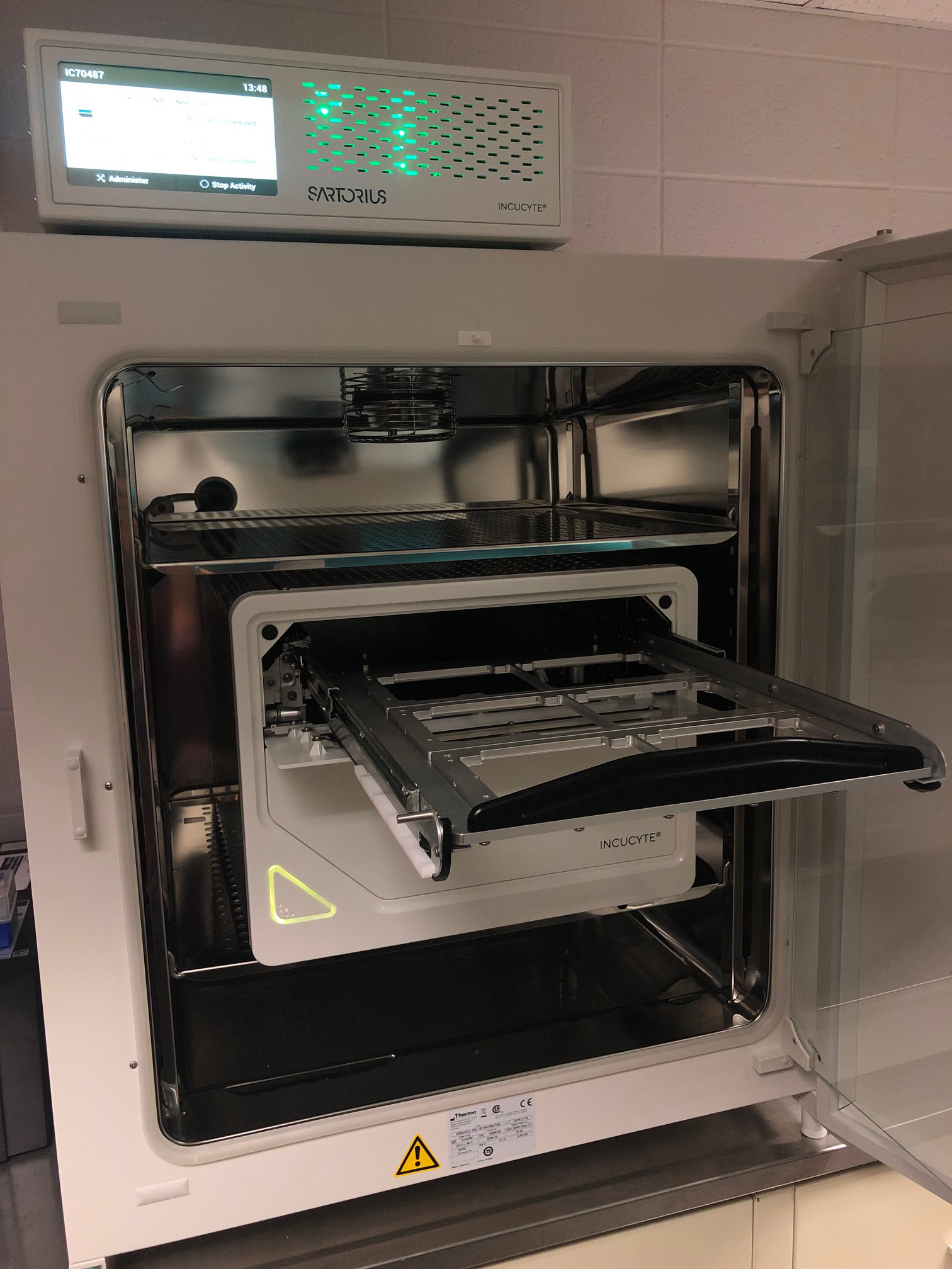
Sartorius Incucyte Live Cell Imaging System
The Incucyte Live Cell Image Real-Time Analysis system is housed in a dedicated incubator, allowing multiple-day experiments, tracking up to 3 fluorophores and a phase image (green, orange and NIR). We have two versions of the Incucyte, tthe SX5 (located in the Hudson Weber Building, room 615) as well as a slightly older model, the SX3, located in Scott Hall, room 6339. The specs for each system are as follows:
SX5: Green excitation 453-485 nm, emission 494-533 nm; Orange excitation 546-568 nm, emission 576-639 nm; NIR excitation 648-674 nm, emission 685-756 nm
SX3: Green excitation 441-481 nm, emission 503-544 nm; Red excitation 567-607 nm, emission 622-704 nm
Both systems have 4x, 10x, and 20x objectives provide varying levels of microscopic analysis.
A robust, user-friendly interface is accessible in our Hudson Weber or Scott Hall lab or from the PI's own computer, allowing remote control of the experiment as it occurs in real time.
Extensive data analysis options provide powerful visualization of images and kinetic measurements.
Schedule an appontment on the Incucyte.
